Intraoperative Neurosurgical Ultrasound — Image Gallery
Overview
This article presents intraoperative neurosurgical ultrasound images from two clinical cases: brain tumor resection and ventricular catheter placement. All images were acquired using high-frequency intraoperative transducers with real-time guidance.
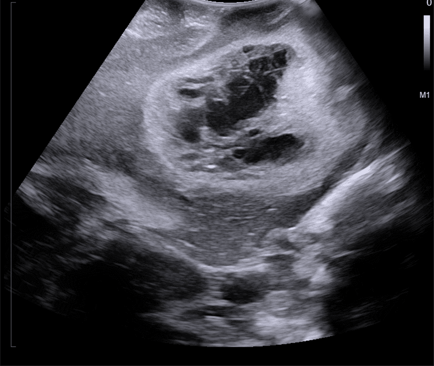
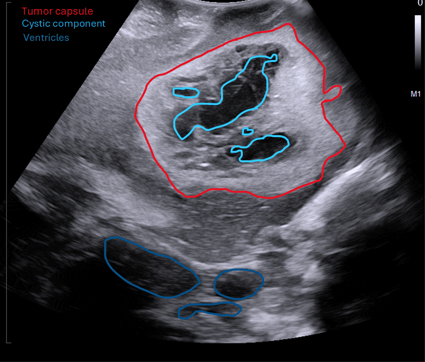
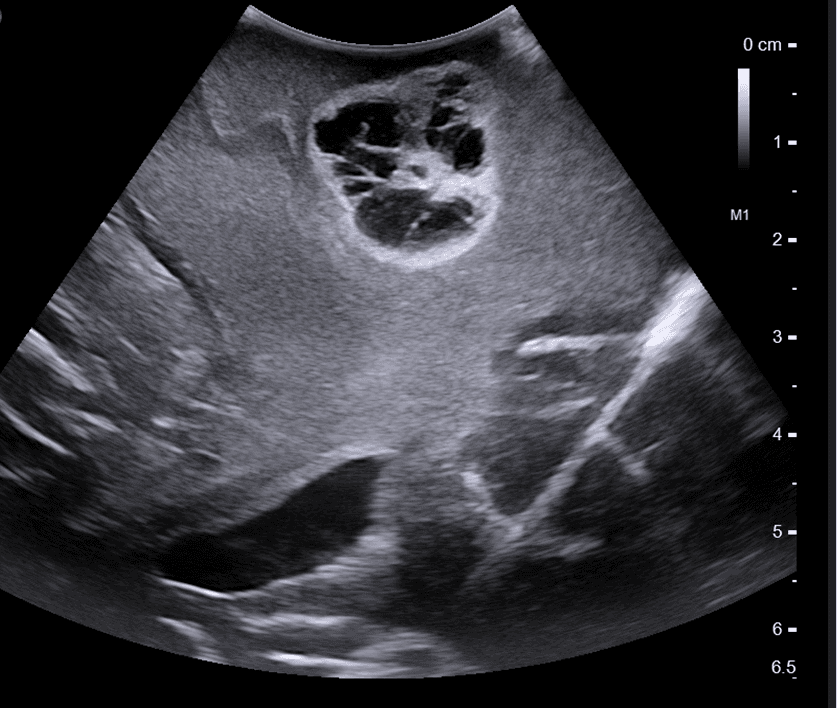
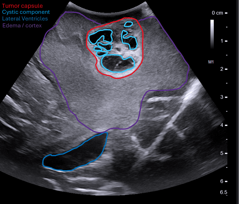
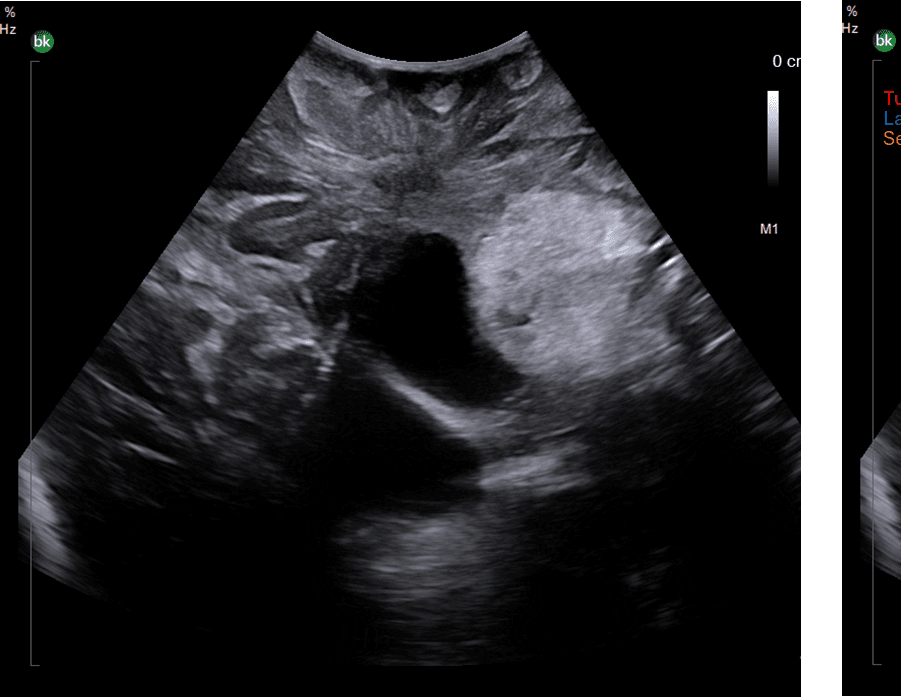
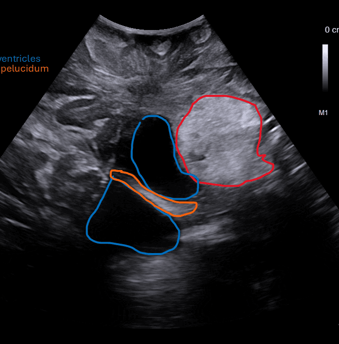
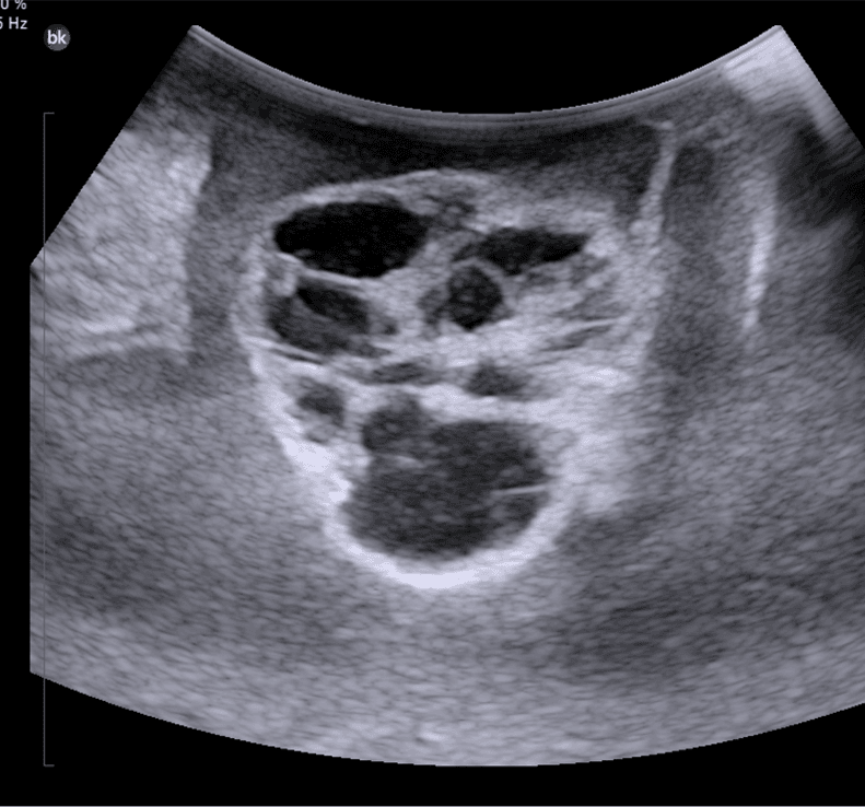
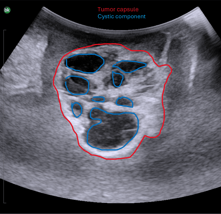
Brain Tumor — Intraoperative Ultrasound
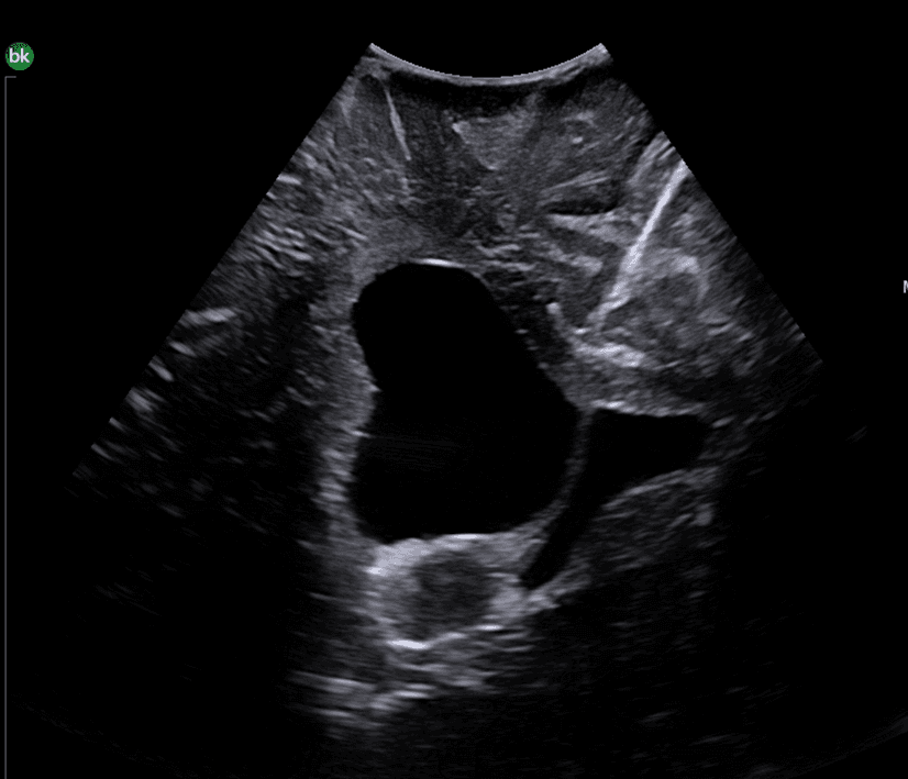
Intraoperative ultrasound is an essential tool for real-time localization of brain tumors during resection. The images below demonstrate key anatomical landmarks including the tumor capsule, cystic components, lateral ventricles, and surrounding cortex.
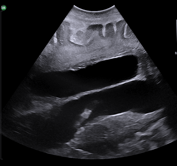
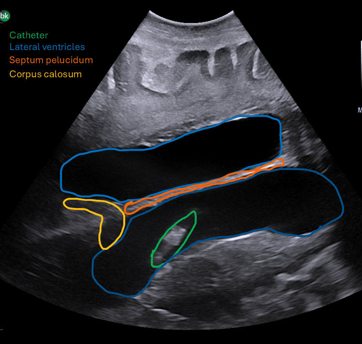
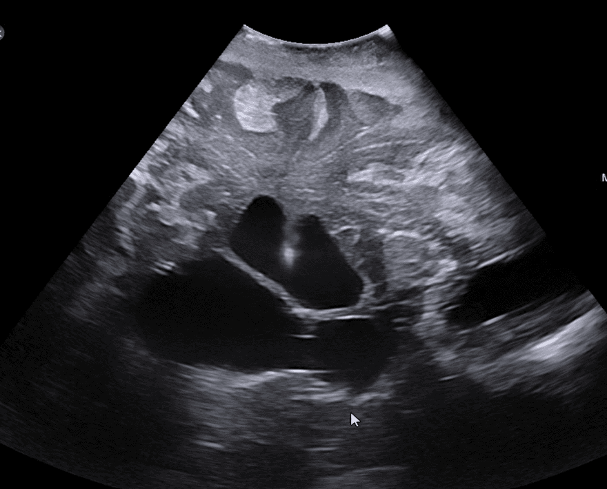
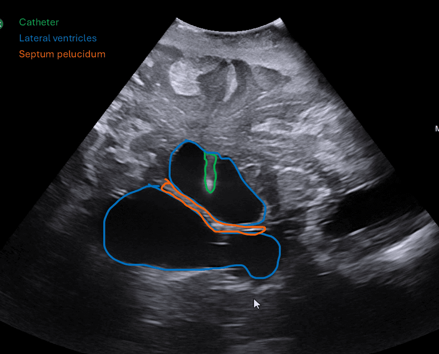
Ventricular Catheter Placement
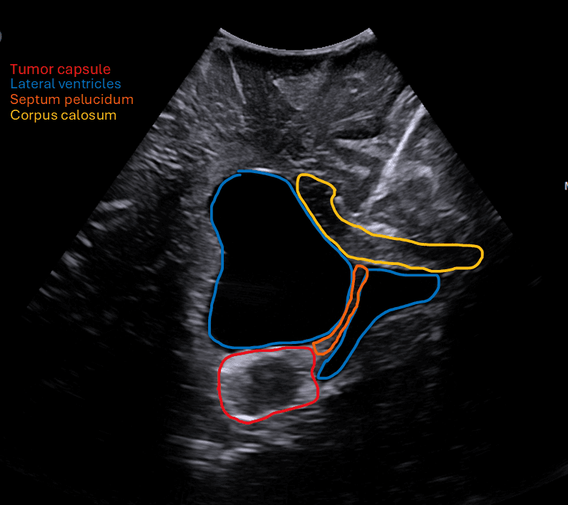
Ultrasound guidance for ventricular catheter (VP shunt) placement provides real-time visualization of catheter trajectory relative to the lateral ventricles, septum pellucidum, and corpus callosum, significantly reducing misplacement rates.
Technique
Images acquired using intraoperative B-mode ultrasound with a high-frequency transducer (7.5–15 MHz) through a craniotomy window. All structures are labeled per standard neurosurgical ultrasound nomenclature.
Image Gallery